-
×
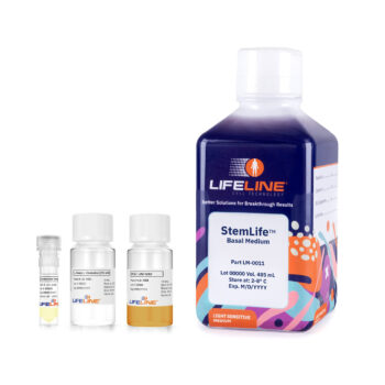
 StemLife MSC Medium Complete Kit
1 × $182.60
 -
×
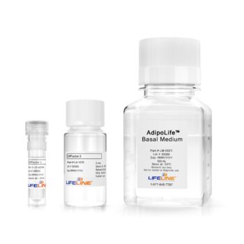
 AdipoLife ™ DfKt ™-1 Adipogenesis Medium Kit
1 × $191.60
Subtotal: $374.20

Mesenchymal stem cells (MSCs) originate from the mesoderm during embryonic development and give rise to multiple different tissue types including bone, cartilage, and fat. It is important to note that while the embryonic mesenchyme produces both connective tissue and hematopoietic tissue types, MSCs do not differentiate into hematopoietic cells.
MSCs are typically isolated from bone marrow, however, they can also be obtained from other tissues including blood (peripheral or cord), placenta, fat, and muscle. One particularly rich source of MSCs is the umbilical cord. Also known as Wharton’s Jelly, umbilical cord tissue has a higher concentration of MSCs than the umbilical cord blood and has the advantage of being an easily obtainable tissue source.
Human MSCs are a robust stem cell culture model:
MSCs provide ample opportunity to conduct studies on basic biology, development, stem cell biology, stem cell differentiation, and cell fate.
Differentiated cell types derived from normal human MSCs also provide a healthy source of cells or tissue that can be used to study various debilitating diseases such as osteoporosis (bone loss), osteoarthritis (cartilage loss), sarcopenia (muscle wasting), obesity, diabetes, inflammatory diseases, and cancer.
Lifeline Cell Technology® has just what you need for your studies involving human MSCs and their cell derivatives. We offer multiple normal human MSC (HMSCs) derived from different tissues as well as HMSC culture media, stem cell differentiation kits, and cell staining kits.
Lifeline provides the following Human Mesenchymal Stem Cells from different tissue sources:
Lifeline® HMSCs are cryopreserved to ensure optimal stem cell phenotype, morphology, and the highest viability and plating efficiency.
Our HMSCs are quality tested via flow cytometry to ensure proper expression of multiple markers of mesenchymal stem cells.
Our HMSCs are uniformly positive (>95%) for integrin CD29, matrix receptors CD44 and CD105, and stromal cell-associated markers CD73, CD90, and CD166.
Additionally, HMSCs are uniformly negative (<2%) for hematopoietic lineage markers CD14, CD31, CD34, and CD45.
HMSCs can be expanded in an undifferentiated state using the following:
These three media formulations are offered as Complete Medium Kits that come with our StemLife MSC basal medium and StemLife MSC LifeFactors specific for each MSC line. For your convenience, all of our StemLife Medium Kits are prepared without phenol red or antimicrobials to provide ideal culture conditions that will not influence experimental outcomes or affect cell viability or function.
Lifeline® HMSCs can be efficiently differentiated into the typical mesenchymal lineages using our tailored differentiation medium kits (ChondroLife, OsteoLife, AdipoLife 1 and AdipoLife 2).
When using Lifeline® HMSCs and stem cell differentiation kits, HMSCs show superior differentiation in comparisons with competing products.
Lifeline® also provides staining kits (Alcian Blue, Alizarin Red, and Oil Red O) to characterize your differentiated cell types following use of our MSC differentiation kits. Check out the table below to see a summary of our HMSC products:
Summary of Lifeline® HMSC Products
| CELL TYPE | HMSC Differentiation Kits | Staining Kits |
| Chondrocytes | ChondroLife Chondrogenesis Medium | Alcian Blue (sulfated proteoglycan deposits) |
| Osteocytes | OsteoLife Osteogenesis Medium | 2% Alizarin Red (calcium deposits) |
| Adipocytes | AdipoLife DfKt-1 Adipogenesis Medium (HMSC-Ad, HMSC Pre-Adipocytes), AdipoLife DfKt-2 Adipogenesis Medium (HMSC-Ad, HMSC-BM, and HMSC-WJ) | Oil Red O (lipids) |
Numerous researchers rely on our high quality normal human MSCs, optimized MSC media, and differentiation kits for their scientific studies. Here are a few highlights from recent publications:
Lifeline® has got you covered when it comes to mesenchymal stem cell research. You can conduct entire studies using our normal HMSCs, MSC differentiation kits, and cell staining kits. Let us know what you’re working on.
References:
2) Prugh et al.