
Fibroblasts: Oh, the Places You Will Go . . .
What are Fibroblasts?
Fibroblasts are connective tissue cells that secrete the components that make up the extracellular matrix and the stroma. Both these structures provide a supportiv framework that keeps cells, tissues, and organs in their proper places.
There are many different types of fibroblasts located in organs and tissues throughout the body. The main structural protein secreted by fibroblasts is collagen; however, others include: glycoproteins, glycosaminoglycans, and various fibers.
Fibroblasts are well known for their role in wound healing. When injury occurs, fibroblasts near the injury site will proliferate, travel to the site of injury, and secrete collagens and other proteins to isolate the injury and facilitate wound repair.
Other members of the connective tissue family include bone, cartilage, fat, and smooth muscle cells. Fibroblasts are particularly interesting, as they are believed to have the ability to convert into other connective tissue cell types when exposed to certain culture conditions.
What You Can Study Using Fibroblasts
Fibroblasts provide an ideal cell model system to study various biological events as well as disease states. Here are a few examples of the types of studies you can conduct on fibroblasts:
- Cell growth and differentiation
- Wound healing
- Skin diseases
- Cancer
- Drug toxicology or screening
- Gene delivery or genome editing
Lifeline® Has the Tools You Need
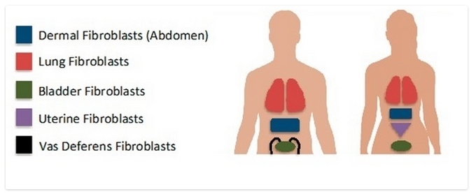
To facilitate your research, Lifeline® provides normal human fibroblasts from the following tissues and organs:
- Bladder
- Lungs (airway)
- Reproductive system (vas deferens and uterine)
- Skin (neonatal and adult)
All of our fibroblast cells should be cultured in our special FibroLife® cell culture medium to achieve optimal cell growth, proliferation, and morphology. To cater to specific experimental needs, we offer different variations of our original FibroLife media including:
- FibroLife® serum free medium
- FibroLife® S2 (low serum – 2%)
- FibroLife® Xeno-Free (which contains no animal-derived components)
Where You Can Go with our Fibroblasts
At least 19 publications have used Lifeline® fibroblast cells or fibroblast culture media (see our References page for the list). Let’s take a look at a few of these studies to see how our fibroblasts and cell culture products were used.
New discoveries in tissue engineering
Davis et al. developed protein polymer hydrogels that closely mimic the function of the extracellular matrix. They were able to culture Lifeline® primary neonatal human fibroblasts in both 2D and 3D hydrogel culture systems using our FibroLife® S2 medium and reported over 95% cell viability. Furthermore, the authors were able to modify the properties of their hydrogels by altering the protein polymer compositions to include bioactive components that directly affect cell function. These interesting findings can be used for tissue engineering applications and improving in situ therapies.
Oligonucleotide therapy accelerates wound healing
Yamanoto et al. found that immunostimulatory CpG oligodeoxynucleotide (ODNs) accelerate wound healing in primates. These ODNs were engineered to target and activate the Toll-like receptor 9 (TLR9), which stimulates immune cells to repair wounds. When skin lesions biopsies from rhesus macaques were treated with a topical gel containing ODNs, wound healing was significantly faster compared to control treatments. To further prove the effectiveness of ODN treatment for wound healing, the authors treated Lifeline® adult human dermal fibroblasts cultured in Fibrolife basal medium with ODNs. They observed increased production of the bFGF (a growth factor that facilitates fibroblast proliferation and wound healing) in fibroblasts treated with ODNs.
Wise advice from Dr. Seuss
Now you know that the places your experiments can go with our fibroblast cells and products are endless. So we will leave you with these words of encouragement from our childhood friend Dr. Seuss!
“You’re off to Great Places! Today is your day! Your mountain is waiting, So… get on your way!”