
New Model System to Evaluate Subcutaneous Drug Absorption
Skin Cancer Awareness Month
Skin cancer, including melanoma, is the most common cancer in the United States—but also one of the most preventable. Practicing sun safety and prioritizing early detection are essential steps in protecting skin health. As the body’s largest organ and first line of defense, the skin plays a vital role not only in preventing disease, but also in how therapeutics are delivered. For protein-based therapies, subcutaneous delivery is often favored due to its accessibility, reduced clinical costs, and potential for sustained-release formulations. To support the development of these therapeutics, physiologically relevant preclinical models are needed to accurately characterize drug absorption in the subcutaneous space.
At Lifeline Cell Technology, our primary human skin cells and specialized media are used by global researchers to uncover new insights across cancer biology, wound healing, drug development, and more. Our catalog includes:
- Human Dermal Fibroblasts – Neonatal, Primary
- Epidermal Keratinocytes — Neonatal, Primary
- Epidermal Melanocytes — Neonatal
- Epidermal Melanocytes — Neonatal, Primary
- Normal Human Dermal Fibroblasts — Adult, Primary
- Epidermal Keratinocytes — Adult, Primary
- Epidermal Melanocytes — Adult
- Human Dermal Fibroblasts – Neonatal, Xeno-Free, Primary
- Human Epidermal Keratinocytes, 10-Donor Pool
- Epidermal Melanocytes — Neonatal, Highly Pigmented
- Epidermal Melanocytes — Adult, Highly Pigmented
- Human Oral Keratinocytes (Gingiva)
- FibroLife S2 Fibroblast Medium Complete Kit
- FibroLife Fibroblast Serum Free Medium Complete Kit
- DermaLife K Keratinocyte Medium Complete Kit
- DermaLife M Melanocyte Medium Complete Kit
- DermaLife Ma Melanocyte Medium Complete Kit
- DermaLife M Melanocyte Medium Complete Kit
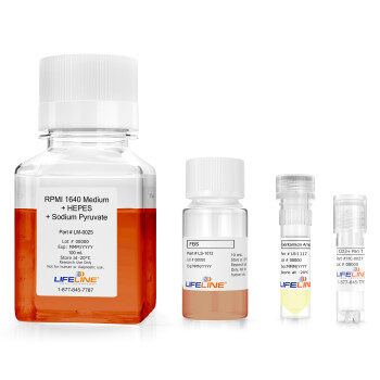
In our feature publication, Offeddu et al. use Lifeline®’s primary dermal microvascular endothelial cells (ECs) and primary dermal fibroblasts (FBs), cultured in VascuLife Endothelial Medium and FibroLife Fibroblast Medium, respectively, as the cellular components to establish their model system, which mimics the subcutaneous microenvironment. This provides a more physiologically relevant mechanism to evaluate absorption and bioavailability of subcutaneously delivered drugs.
Microphysiological Models of the Subcutaneous Space for Preclinical Development of Subcutaneously Delivered Protein Therapeutics
As subcutaneous delivery continues to gain traction for protein-based therapies, a critical gap remains in accurately predicting subcutaneous bioavailability in humans during preclinical development. Current models often lack the fidelity needed to accurately replicate the human subcutaneous space. To address this, Offeddu and colleagues have developed a novel microphysiological model that mimics the human subcutaneous environment, enabling direct measurement of key parameters, such as interstitial transport and vascular absorption, which influence drug bioavailability.
Human dermal endothelial cells, fibroblasts, and adipocytes were embedded in a 3D fibrin matrix within a microfluidic device to create the microphysiological hypodermal model. Over 5–7 days, the cells self-assembled into a perfusable, interconnected microvascular network that closely replicates the architecture of the human subcutaneous (hypodermal) space.
Morphological comparison and immunofluorescent staining (CD31 and podoplanin) of fixed human skin biopsies show that the dynamic microvascular network (dMVN) model can replicate key vascular features of human skin. While the dMVNs exhibited greater vascular density and larger average vessel diameters (~5 μm), their comparable specific surface area to the native sub-epidermis and hypodermis makes them functionally relevant for predicting subcutaneous drug absorption.
Endothelial and Interstitial Transport
The dMVN microfluidic model can be used to assess the impact of molecular physicochemical characteristics on protein transport. The researchers evaluated how protein size, concentration, and charge affect basolateral-to-apical transport across the endothelial barrier. Consistent with known biology, the model showed that large proteins like IgG move from the tissue into the bloodstream via transcytosis, a saturable mechanism where higher IgG concentrations reduce transport efficiency. They also found that molecular charge influenced diffusion in the ECM, with positively charged dextran diffusing less readily than neutral or negatively charged molecules. The diffusivity values obtained in the dMVN model for a diverse set of globular proteins (5.8-150 kDa) align with previously published reports from animal models. These findings suggest that endothelial permeability and protein–ECM interactions in the model closely mirror in vivo behavior, making the model a valuable tool for understanding subcutaneous drug absorption.
Lymphatic Transport
Protein therapeutics injected subcutaneously can be absorbed through both the blood microvasculature and the lymphatic system. The dMVN model was compared to existing lymphatic microvascular models, revealing that for larger proteins like serum IgG, absorption through the lymphatic endothelium is favored over direct entry into the bloodstream. Permeability tests for microvascular and lymphatic serum IgG showed significantly higher rates of entry through the lymphatic system at every IgG concentration tested. The lymphatic absorption process was also found to be largely non-selective, in contrast to the size-dependent transport seen in the blood microvasculature. These findings suggest that the lymphatic network plays a predominant role in the absorption of monoclonal antibodies following subcutaneous administration.
An essential step in preclinical drug development is predicting human subcutaneous bioavailability, a pharmacokinetic outcome that is highly variable and difficult to predict with current models. The dMVN model described in this study provides a unique tool to measure biophysical parameters within the hypodermal microenvironment, helping to better understand protein kinetics and distribution at the injection site. This approach is valuable for optimizing the development of protein therapeutics intended for subcutaneous administration with optimal bioavailability.
Join us next month for another installment of the Lifeline® blog to see how our cells and culture media are advancing biomedical research worldwide. If you have used our products in your publication, we’d love to feature your work here!